
Google Earth for the Brain
If BrainMaps.org is like Google Maps for the Brain, StackVis is Google Earth for the Brain. Welcome to StackVis, a 3D viewer of neuroanatomical sections.
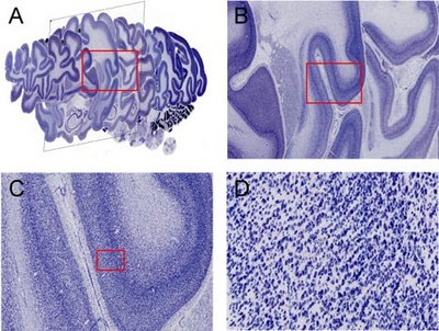
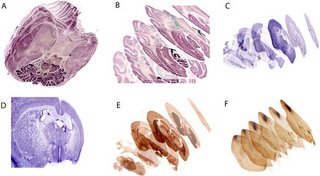 Figure Caption. The development of additional desktop application tools for interacting with brainmaps.org image and database data includes the one shown here, StackVis, which is a 3D viewer of neuroanatomical virtual slide image stacks that is integrated with high resolution viewing of and interaction with individual sections comprising the image stack. (A) horizontal image stack of nissls of the macaque brain viewed from below. (B) same image stack as in (A) but from a different perspective, with increased inter-section spacing, and with areal and nuclear labels. (C) coronal image stack of nissls of the mouse brain. (D) a section from (C) viewed at higher resolution. (E) coronal image stack of acetylcholinesterase reacted sections of the mouse brain. (F) sagittal image stack of the mouse brain reacted for biotinylated dextran amine (BDA) following an injection in frontal cortex. Note that labeled fibers can be followed within the image stack due to section transparency and that individually labeled subcortical structures can be discerned, allowing for an assessment of labeled fiber pathways and areas within a 3D framework. In addition, StackVis features automated section registration and edge detection capabilities.
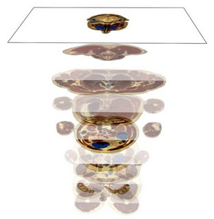 Figure Caption. Viewing the Visible Male using StackVis. The sections are axial, and are arranged in this figure so that the brain is located at the top and the legs are located at the bottom of the image stack.
Labels: 3D brain, brain atlas, brain mapping, google earth for brain
posted by neubrain at 11:08 PM
![]()
![]()
0 Comments:
Post a Comment
<< Home