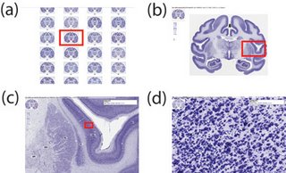
Virtual microscopy is a method of posting microscope images on, and transmitting them over, computer networks. This allows independent viewing of images by large numbers of people in diverse locations.
Prior to recent advances in virtual microscopy, slides were commonly digitized by various forms of film scanner and image resolutions rarely exceeded 5000 dpi. Nowadays, it is possible to achieve more than 100,000 dpi and thus resolutions approaching that visible under the optical microscope. This increase in scanning resolution comes at a price; whereas a typical flatbed or film scanner ranges in cost from $200 to $600, a 100,000 dpi slide scanner will range from $80,000 to $200,000.
Virtual microscopy has been characterized as potentially a
disruptive technology. A disruptive technology is a new technological innovation, product, or service that eventually overturns the existing dominant technology in the market, which in this case would be real (i.e., conventional) microscopy. Our experience with virtual microscopy suggests that it is unlikely to replace real microscopy any time soon, but for the time being, it nicely complements and extends the capabilities of real microscopes. Specifically, we find a three-fold extension of virtual microscopy over real microscopy in the following areas: 1) data-sharing and remote access, 2) data-management and annotation, and 3) data-mining. Data management and data-mining of virtual (digitized) slides are capabilities that cannot be directly applied to real slides. In addition, the online distribution and sharing of virtual slides with anyone with an internet connection ensures the rapid dissemination of neuroanatomical data that otherwise would not be possible. While largely emphasizing the pros of virtual slides in this article, it is worthwhile to point out the cons. Namely, it is not possible to change focus in a virtual slide as it is in a real slide. Normally, this is not a problem since virtual slides tend to be completely in focus. However, the inability to change the plane of focus in a virtual slide rules out their use in unbiased stereological estimation methods that require optical dissectors. Nonetheless, biased sterelogical estimation methods, or unbiased methods not using optical dissectors, are still possible. Another drawback is that the resolution of the virtual slide is limited to the optical lens used in the scanner. For example, if we generate a virtual slide at 20x and subsequently want to examine part of the slide at 40x, then it is necessary to rescan the entire slide using the higher objective, which in some cases, is not possible due to file size restrictions or hardware issues. Finally, at the time of writing, virtual microscopy does not deal well with fluorescence, and is only recommended for light microscopy.
Virtual microscopy-based digital brain atlases are superior to conventional print atlases in five respects: 1) resolution, 2) annotation, 3) interaction, 4) data integration, and 5) data-mining. The resolution of conventional print brain atlases typically does not exceed 7200 dpi, whereas virtual microscopy-based digital brain atlases attain 100,000 dpi and offer the ability to zoom in and out. Annotation can be more complete in virtual microscopy-based digital brain atlases, with options to display some types of annotations and make the rest invisible. Greater interactivity means that the user can zoom in/out and pan through brain image data, which is not possible in print-based atlases. Data-integration capabilities, including the integration of connectivity and gene expression data, are superior for the virtual microscopy-based digital brain atlases. And finally, the ability to data-mine virtual microscopy-based digital brain atlases is a feature not available for print-based atlases.
While we do not foresee virtual microscopy-based digital brain atlases completely replacing conventional print-based brain atlases, we expect that they will be progressively more commonly used in place of print-based atlases.What I'd like to know is, how likely is virtual microscopy to overtake regular microscopy and print atlases in the near future? In my opinion, conventional print brain atlases are a ripoff, often costing over $200 each. The idea of having this information freely available online is very tantalizing and welcome.
In other news,
Society for Neuroscience 2006 is fast approaching! Hope to see you all in Atlanta this Oct 14-18.
Labels: brain atlas, brain mapping, virtual microscopy


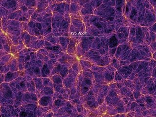
 Does the large-scale structure of the universe resemble a big neural net? Yes, I know it sounds somewhat ridiculous, but I couldn't help making the association when I saw these pictures of the large-scale structure of the universe. What would be the implications of a universe-wide intelligence made explicit in the large-scale structure of the universe and its interactions?!